The Hidden Highway of Sensation: How the Cervical Plexus Governs Our Neck and Beyond
The Hidden Highway of Sensation: How the Cervical Plexus Governs Our Neck and Beyond
Often operating in the shadow of its more famous counterparts, the cervical plexus is the intricate nerve network responsible for the quiet, constant language of feeling in our neck, head, and diaphragm. This complex system, formed by the intricate weaving of the first four cervical nerves, translates the harsh sting of a winter wind on our skin and the subtle pressure of a collar button into signals our brain can understand. Understanding this neural architecture is not merely an academic exercise; it is fundamental to diagnosing and treating a wide array of neurological and orthopedic conditions that impact daily comfort and motor function.
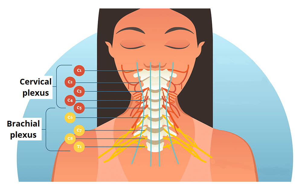
Unlike a single, discrete nerve, the cervical plexus is a sophisticated convergence point where the ventral rami of spinal nerves C1 through C4 merge. This anatomical crossroads gives rise to two primary branches: the cutaneous branches, which form the supraclavicular, transverse cervical, and lesser occipital nerves, mapping sensation across the scalp, neck, and shoulder; and the muscular branches, which include the phrenic nerve, the sole motor driver of the diaphragm. The result is a sophisticated sensory and motor map that governs both our awareness of the world around our head and the life-sustaining rhythm of our breathing.
The Anatomical Architecture of a Neural Network
The cervical plexus is not a static structure but a dynamic network deeply embedded in the musculature of the neck. Its formation begins in the neck’s posterior triangle, a region bounded by the sternocleidomastoid muscle in front, the trapezius muscle behind, and the clavicle below. Here, the nerves interlace in a pattern that can vary slightly from person to person, creating a unique anatomical fingerprint for each individual. This variability is a key consideration for clinicians, as it explains why nerve blocks might have slightly different effects from one patient to the next.
Structurally, the plexus can be broken down into its major functional pathways:
- The Cutaneous (Sensory) Branches: These nerves fan out to provide sensory innervation to specific dermatomes, or patches of skin.
- The Muscular (Motor and Sensory) Branches: These supply the muscles of the neck and back, as well as the critical phrenic nerve.
- The Communicating Branches: These facilitate dialogue with other nerve networks, such as the vagus nerve, integrating the cervical region with broader systemic functions.
The Sentinel of Sensation: Cutaneous Innervation
The sensory branches of the cervical plexus act as the body’s security system for the upper torso and head. The lesser occipital nerve, arising from the second cervical nerve (C2), provides sensation to the skin behind the ear and the upper part of the neck. Damage or irritation to this nerve can manifest as a persistent, burning pain or numbness, often misdiagnosed as a simple headache or ear infection.
Moving down the chain, the transverse cervical nerve (C2, C3) covers the front of the neck, while the supraclavicular nerves (C3, C4) extend over the shoulder and down the upper chest. These nerves are frequently the culprits in “referred pain” syndromes. For instance, a problem in the cervical spine can sometimes project discomfort directly across the supraclavicular region, creating a confusing diagnostic picture for physicians.
“The cervical plexus is a prime example of how anatomy dictates symptomology,” explains Dr. Aris Thaler, a neurologist at the Institute for Advanced Neural Mapping. “When a patient presents with burning pain along the jawline and neck, we don't just look at the spine; we map the dermatomes. More often than not, we are tracing the path of the cervical plexus’s cutaneous branches to find the root of the issue.”
The Breath of Life: The Phrenic Nerve and Motor Function
While sensation is a critical function, the cervical plexus’s most vital role is arguably its control of the diaphragm via the phrenic nerve. Originating predominantly from the C4 nerve root—leading to the memorable medical mnemonic “C3, 4, 5 keeps the diaphragm alive”—the phrenic nerve is the sole motor supply to this crucial muscle. Each phrenic nerve sends signals that cause the diaphragm to contract and descend, creating the negative pressure necessary for inhalation. Without this constant, unconscious signaling, breathing would cease.
Beyond respiration, the muscular branches supply the muscles of the neck (including the sternocleidomastoid and trapezius), allowing for the complex movements of head rotation, flexion, and shoulder shrugging. Injury to these branches can result in a characteristic “shoulder drop” or an inability to turn the head against resistance, profoundly impacting a person’s quality of life.
Clinical Pathologies and Modern Diagnostics
When the cervical plexus is compromised, the symptoms can be both diverse and debilitating. One of the most common iatrogenic (physician-caused) injuries is a phrenic nerve palsy, which can occur as a complication of cardiac surgery or central line placement. This condition can lead to shortness of breath, particularly when lying flat, as the compromised diaphragm struggles to function.
Other pathologies include:
- Cervical Plexus Neuropraxia: Often resulting from a sudden, forceful neck stretch (as in a car accident), this “burning neck syndrome” causes severe, shooting pain and sensory disturbances.
- Thoracic Outlet Syndrome: While primarily involving the brachial plexus, compression in the thoracic outlet can impinge on the lower cervical nerves, affecting the plexus.
- Iatrogenic Injury: Procedures such as cervical lymph node biopsies or carotid artery stenting carry a risk of damaging the surrounding nerve network.
Diagnosing issues within the cervical plexus relies heavily on a combination of clinical examination and advanced imaging. While a standard MRI can show structural issues in the spine, more specific tests are often required. Electromyography (EMG) and nerve conduction studies are the gold standard, allowing physicians to electrically stimulate specific branches of the plexus and measure the response, pinpointing the exact location and severity of the lesion.
The Frontier of Treatment and Research
Treatment for cervical plexus disorders is as varied as the symptoms themselves. For acute inflammation, corticosteroids and physical therapy are often the first line of defense. For chronic, intractable pain, however, more advanced interventions are required.
One of the most promising developments in the field is the use of targeted nerve blocks. Using advanced imaging guidance, physicians can inject anesthetic directly at the point of irritation, providing both diagnostic confirmation and immediate relief. For patients with permanent nerve damage, surgical options such as neurolysis or even nerve grafting are being explored to restore function.
Looking to the future, research into neurostimulation offers a glimpse of a new horizon. Scientists are investigating devices that can modulate the activity of the cervical plexus, potentially offering relief for conditions like chronic headaches and certain types of tinnitus that have a cervical origin. As our understanding of this hidden highway deepens, the potential to alleviate suffering and restore quality of life for millions of patients becomes increasingly tangible.
Related Post
Unraveling the Mysteries of the Cervical Plexus of Nerves: Understanding its Anatomy and Function

Unlock the Secrets of Your Neck: Understanding the Cervical Plexus

The Mysterious World of Mail From Po Box 6753 Sioux Falls Sd: Uncovering the Truth Behind the Enigmatic Envelopes

Latest Update: Kris Marshall in Critical Condition After Horrific Car Accident - Fans and Friends Rally Around


