The Optic Canal: Unraveling the Critical Gateway for Vision and Its Hidden Dangers
The Optic Canal: Unraveling the Critical Gateway for Vision and Its Hidden Dangers
The optic canal is far more than a mere anatomical footnote; it is a vital bony corridor that serves as the exclusive pathway for the optic nerve and ophthalmic artery, acting as the essential bridge between the eye and the brain. Understanding its structure, function, and the clinical implications of its compromise is fundamental to neurology, ophthalmology, and neurosurgery. This article explores the intricate anatomy, physiological significance, and the serious conditions that arise when this critical channel is disrupted.
The human skull is a complex fortress of bone, designed to protect the delicate organs of sight and thought. Within this intricate architecture, small openings called foramina act as strategic gates, allowing crucial neurovascular structures to travel from the brain to the face and neck. The optic canal, located within the lesser wing of the sphenoid bone, is one of the most significant of these foramina. It is the dedicated tunnel through which the optic nerve (cranial nerve II) and the ophthalmic artery make their perilous journey from the orbit to the cranial cavity. Any pathology that narrows, blocks, or damages this canal directly threatens the precious cargo it carries—our vision.
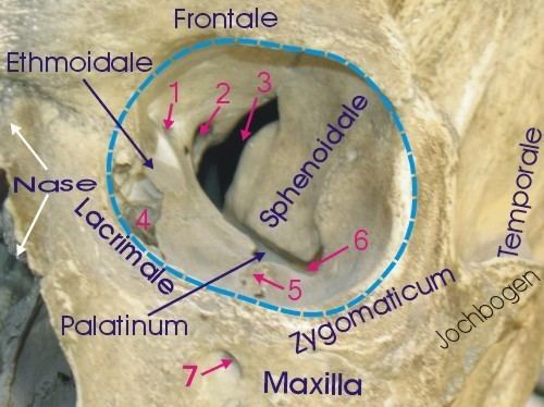
Anatomically, the optic canal is a relatively short, bony passage measuring approximately 10 to 12 millimeters in length. Its entrance, known the orbital or intraorbital opening, is situated in the apex of the orbit, just behind the orbital plate of the ethmoid bone. The canal then traverses medially, forward, and slightly upward before exiting through the superior orbital fissure, although it is specifically demarcated as the optic canal before this wider fissure. The boundaries of this canal are rigid and unforgiving. The medial wall is formed by the body of the sphenoid bone, while the lateral wall is comprised of the lesser wing of the same bone. The superior aspect is defined by the planum sphenoidale, and the inferior boundary is the dorsum sellae and the posterior clinoid process. This bony enclosure provides structural stability but offers little room for expansion, making the structures within highly vulnerable to even minor forms of compression.
The primary cargo of the optic canal is the optic nerve itself, which is actually an extension of the diencephalon rather than a true peripheral nerve. Alongside this neural structure travels the central retinal artery, a critical vessel that supplies the inner layers of the retina. As Dr. Robert M. Scult, an orbital surgeon at the Massachusetts Eye and Ear Infirmary, explains, "The relationship between the optic nerve and the canal is one of intimate confinement. There is virtually no loose tissue within this space, which means any swelling, be it inflammatory, vascular, or neoplastic, creates an immediate and serious threat to the conduit." This intimate anatomy is the reason pathologies affecting the canal often present with rapid and severe visual deficits.
The clinical significance of the optic canal cannot be overstated, as its compromise leads directly to a specific and debilitating syndrome. **Optic Canal Syndrome**, also known as Optic Nerve Compression, manifests through a distinct set of symptoms that progress with the severity and duration of the compression. The most profound symptom is visual loss, which can range from a subtle decrease in visual acuity to total blindness. This is frequently accompanied by a diminished or absent direct pupillary response (afferent pupillary defect) and a loss of color vision, particularly for reds and greens. Pain, especially with eye movement, may also be a presenting feature due to the inflammation or stretching of the surrounding dural sheaths.
Several pathological processes can lead to this syndrome, each with its own mechanism of compression:
1. **Tumors:** This is one of the most common causes. The differential diagnosis includes meningiomas, which often arise from the meninges surrounding the canal, as well as metastases, pituitary adenomas that extend upward, and olfactory groove tumors. These slow-growing masses exert a gradual but relentless pressure on the nerve.
2. **Inflammatory Conditions:** Diseases such as sarcoidosis, Tolosa-Hunt syndrome (a painful granulomatous inflammation), and autoimmune vasculitis can cause swelling and granuloma formation within the confined space of the canal.
3. **Trauma:** A fracture of the lesser wing of the sphenoid bone can directly lacerate or contuse the optic nerve. Additionally, post-traumatic bleeding or edema can fill the canal, leading to acute compression.
4. **Vascular Abnormalities:** An aneurysm of the internal carotid artery, particularly at the junction of the posterior communicating artery, can expand and impinge on the nerve running alongside the canal.
5. **Idiopathic Intracranial Hypertension (IIH):** While this condition causes generalized papilledema, the increased intracranial pressure can transmit down the subarachnoid space surrounding the optic nerve as it passes through the canal, leading to secondary compression at this narrowest point.
Diagnosing conditions related to the optic canal relies heavily on advanced neuroimaging. **Magnetic Resonance Imaging (MRI)** with high-resolution sequences is the gold standard. It provides exceptional soft-tissue contrast, allowing clinicians to visualize the optic nerve itself, identify enhancing tumors, track inflammation, and assess blood flow. Specifically, T1-weighted images with fat suppression can help delineate the nerve, while magnetic resonance angiography (MRA) or venography (MRV) can evaluate vascular causes. When MRI is contraindicated or additional detail of the bony anatomy is required, **Computed Tomography (CT) scanning** comes into play. CT scans are superb for visualizing fractures, bony erosions caused by slow-growing tumors, and the precise dimensions of the canal itself.
Treatment is entirely dependent on the underlying cause and the acuteness of the visual threat. For compressive tumors or aneurysms, neurosurgical intervention is often the only recourse. The goals of surgery are to decompress the optic nerve by removing the mass and to restore cerebral spinal fluid flow if hydrocephalus is present. The prognosis for visual recovery is directly tied to the duration and severity of the compression; the longer the nerve is deprived of its blood supply, the less likely vision is to return. In cases of inflammatory etiology, high-dose intravenous corticosteroids are the first line of defense to rapidly reduce edema and inflammation. For conditions like IIH, the focus shifts to managing intracranial pressure through medications like acetazolamide or, in refractory cases, surgical procedures such as optic nerve sheath fenestration, which creates a small window in the dural sheath to relieve pressure on the nerve as it exits the canal.
The ramifications of neglecting pathology in the optic canal are severe. Permanent vision loss is not the only potential outcome. In cases of traumatic injury or aggressive tumors, the optic nerve can atrophy, leading to a permanent, irreversible blindness in the affected eye. Furthermore, because the optic nerve is closely associated with the ophthalmic artery, severe compression can also lead to ocular ischemia, affecting not just the retina but other structures within the eye, potentially causing neovascular glaucoma—a particularly painful and difficult-to-manage complication. Early recognition and intervention are paramount. As one neurologist specializing in skull base disorders puts it, "The optic canal is a window to the brain, but when it becomes a choke point, it becomes a window to a disaster. We must look through it, but we must also protect it."
In summary, the optic canal is a critical anatomical corridor whose integrity is fundamental to the preservation of sight. It is a space where neurosurgical, ophthalmic, and neurologic pathologies converge. From slow-growing meningiomas to acute fractures, the spectrum of threats to this canal is diverse. Through a combination of vigilant clinical examination, sophisticated imaging, and timely intervention, the medical community continues to strive to preserve this vital gateway, protecting one of our most precious senses—the ability to see.
Related Post
The Optic Canal A Critical Yet Often Overlooked Brain Highway

Optic Canal: The Crucial Gateway to Clear Vision and Eye Health


